ستون فقرات
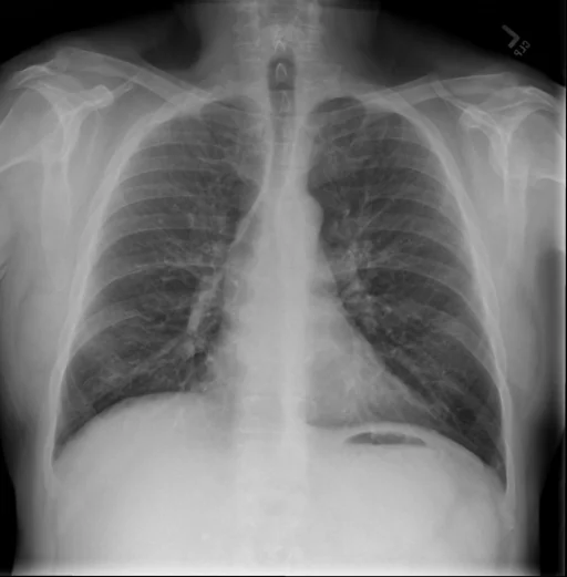
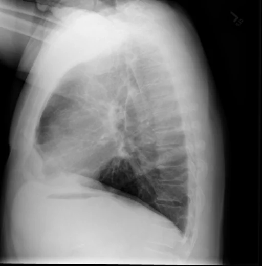
برداشت تشخیصی رادیولوژیست (خلاصهٔ تحلیلی)
یافتههای ثبتشده: ستون فقرات (دژنراتیو) و مهرههای سینهای (دژنراتیو، خفیف). دستهبندی: «ستون فقرات». وضعیت: «فقط یافتهٔ قدیمی/مزمن».
گزارش بالینی اصلی (متن مستند انگلیسی — NoDerivatives)
- اندیکاسیون (Indication)
- Adult male, and preoperative evaluation for right total knee replacement.
- مقایسه با بررسیهای قبلی (Comparison)
- Radiographs of the right knee. PA and lateral views of the chest.
- یافتهها (Findings)
- Chest. Heart size within normal limits. No focal airspace disease. No pneumothorax or effusions. Mild degenerative changes throughout the thoracic spine. Right knee. There has been prior ligamentous repair. There is tricompartmental joint space narrowing and marginal osteophyte formation which is severe in the medial compartment. No knee joint effusion. No fractures or dislocations.
- برداشت نهایی (Impression)
- 1. Chest. No acute cardiopulmonary findings. 2. Right knee. Stable advanced degenerative changes, most severe in the medial compartment.
مشخصات بررسی رادیوگرافی
- شماره پرونده
- CXR ۲۴۵۱
- مودالیتی
- رادیوگرافی قفسهٔ سینه (Chest X-Ray)
- تعداد نماها
- ۲ تصویر
- وضعیت بالینی
- فقط یافتهٔ قدیمی/مزمن
ستون فقرات
این رادیوگرافی قفسهٔ سینه چه یافتهای را نشان میدهد؟
رادیوگرافی قفسهٔ سینه در این پرونده در دستهٔ «ستون فقرات» قرار دارد. یافتهٔ اصلی ثبتشده «ستون فقرات (دژنراتیو)» است و وضعیت مورد «فقط یافتهٔ قدیمی/مزمن» است. اطلاعات تکمیلی: شدت: خفیف.
این متن خلاصهٔ دادههای ساختاریافتهٔ مجموعه است، نه توصیف تصویر.
- شدت و توصیفگران: خفیف
- دستهبندی تشخیصی: ستون فقرات
- پوشش تصویربرداری: ۲ نما، رزولوشن اصلی ۵۱۲×۵۲۱ پیکسل
- کلیدواژههای مرتبط: ستون فقرات، Spine / Vertebrae (degenerative, fusion, osteophyte)، مهرههای سینهای، Thoracic Vertebrae، دژنراتیو، degenerative
کیسهای رادیولوژی مرتبط
منبع، اعتبار علمی و مجوز انتشار
Indiana University Chest X-ray Collection · Open-i / U.S. National Library of Medicine · CC BY-NC-ND 4.0
مجوز انتشار: CC BY-NC-ND 4.0
منبع اصلی داده: https://openi.nlm.nih.gov/