کانژسیون ریوی، تراکم بافتی (دانسیته)، کلسیفیکاسیون ریه
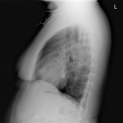
برداشت تشخیصی رادیولوژیست (خلاصهٔ تحلیلی)
یافتههای ثبتشده: کانژسیون ریوی، تراکم بافتی (دانسیته)، کلسیفیکاسیون ریه، کلسیفیکاسیون (ریه، ناف ریه، غدد لنفاوی، چپ، کوچک) و تراکم بافتی (دانسیته) (ریه، لوب میانی، راست، گِرد). دستهبندی: «کلسیفیکاسیون، کانژسیون ریوی و تراکم بافتی (دانسیته)». وضعیت: «یافتهٔ فعال / نامشخص».
گزارش بالینی اصلی (متن مستند انگلیسی — NoDerivatives)
- اندیکاسیون (Indication)
- Sarcoidosis
- مقایسه با بررسیهای قبلی (Comparison)
- With contrast
- یافتهها (Findings)
- There are no focal airspace opacities within the lungs. There is a 1 cm nodular density projecting in the right midlung between the third and fourth right anterior ribs which does not appear to be present on the. To the pulmonary interstitium is not clear, making it the vasculature somewhat indistinct in the mid and lower lungs. This may reflect parenchymal nodules. Nodules were identified on the prior chest CT. Mediastinal contours appear grossly normal. There are small calcified left hilar lymph nodes. The heart and pulmonary vessels otherwise appear normal. Pleural spaces appear clear.
- برداشت نهایی (Impression)
- 1. 1 nodular density seen on the PA view only projecting in the right midlung. Recommend noncontrasted enhanced for evaluation of this nodule. Does this patient have known factors for malignancy? 2. Somewhat indistinct pulmonary interstitium possibly reflecting underlying pulmonary sarcoidosis
مشخصات بررسی رادیوگرافی
- شماره پرونده
- CXR ۲۰۷۱
- مودالیتی
- رادیوگرافی قفسهٔ سینه (Chest X-Ray)
- تعداد نماها
- ۱ تصویر
- وضعیت بالینی
- یافتهٔ فعال / نامشخص
این رادیوگرافی قفسهٔ سینه چه یافتهای را نشان میدهد؟
رادیوگرافی قفسهٔ سینه در این پرونده در دستهٔ «کلسیفیکاسیون، کانژسیون ریوی و تراکم بافتی (دانسیته)» قرار دارد. یافتهٔ اصلی ثبتشده «کانژسیون ریوی» است و وضعیت مورد «یافتهٔ فعال / نامشخص» است. اطلاعات تکمیلی: سمت: چپ و راست، شدت: دوطرفه و گِرد.
این متن خلاصهٔ دادههای ساختاریافتهٔ مجموعه است، نه توصیف تصویر.
- محل درگیری: چپ، راست
- شدت و توصیفگران: دوطرفه، گِرد، کوچک
- دستهبندی تشخیصی: کانژسیون ریوی، تراکم بافتی (دانسیته)، کلسیفیکاسیون
- پوشش تصویربرداری: ۱ نما، رزولوشن اصلی ۵۱۲×۵۱۲ پیکسل
- کلیدواژههای مرتبط: کلسیفیکاسیون، Calcinosis، کانژسیون ریوی، Pulmonary Congestion، تراکم بافتی (دانسیته)، Density
کیسهای رادیولوژی مرتبط
منبع، اعتبار علمی و مجوز انتشار
Indiana University Chest X-ray Collection · Open-i / U.S. National Library of Medicine · CC BY-NC-ND 4.0
مجوز انتشار: CC BY-NC-ND 4.0
منبع اصلی داده: https://openi.nlm.nih.gov/