سایر
برداشت تشخیصی رادیولوژیست (خلاصهٔ تحلیلی)
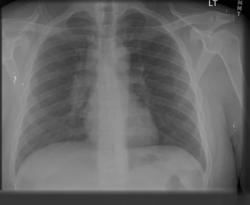
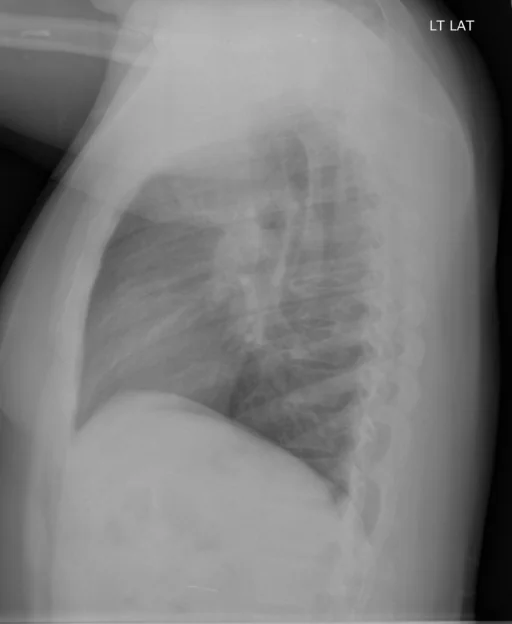
یافتههای ثبتشده: سایر و شریان ریوی (دوطرفه، برجسته). دستهبندی: «سایر». وضعیت: «یافتهٔ فعال / نامشخص».
گزارش بالینی اصلی (متن مستند انگلیسی — NoDerivatives)
- اندیکاسیون (Indication)
- Dyspnea
- یافتهها (Findings)
- The cardiac silhouette and upper mediastinum are within normal limits. There is no pulmonary venous congestion. There is prominence of the pulmonary arteries, right greater than left. There is no acute air space infiltrate, pleural effusion or pneumothorax.
- برداشت نهایی (Impression)
- 1. No acute pulmonary infiltrate or effusion. There is no pneumothorax. 2. Prominent bilateral, right greater than left. This appears slightly increased from the prior studies. Findings could be related to hilar lymph nodes or enlarged pulmonary arteries. If clinically indicated, further evaluation with contrast-enhanced CT of the thorax could be performed.
مشخصات بررسی رادیوگرافی
- شماره پرونده
- CXR ۱۳۸۶
- مودالیتی
- رادیوگرافی قفسهٔ سینه (Chest X-Ray)
- تعداد نماها
- ۲ تصویر
- وضعیت بالینی
- یافتهٔ فعال / نامشخص
سایر
این رادیوگرافی قفسهٔ سینه چه یافتهای را نشان میدهد؟
رادیوگرافی قفسهٔ سینه در این پرونده در دستهٔ «سایر» قرار دارد. یافتهٔ اصلی ثبتشده «شریان ریوی (دوطرفه، برجسته)» است و وضعیت مورد «یافتهٔ فعال / نامشخص» است. اطلاعات تکمیلی: شدت: دوطرفه و برجسته.
این متن خلاصهٔ دادههای ساختاریافتهٔ مجموعه است، نه توصیف تصویر.
- شدت و توصیفگران: دوطرفه، برجسته
- دستهبندی تشخیصی: سایر
- پوشش تصویربرداری: ۲ نما، رزولوشن اصلی ۵۱۲×۴۲۰ پیکسل
- کلیدواژههای مرتبط: سایر، Other، شریان ریوی، Pulmonary Artery، دوطرفه، bilateral
کیسهای رادیولوژی مرتبط
منبع، اعتبار علمی و مجوز انتشار
Indiana University Chest X-ray Collection · Open-i / U.S. National Library of Medicine · CC BY-NC-ND 4.0
مجوز انتشار: CC BY-NC-ND 4.0
منبع اصلی داده: https://openi.nlm.nih.gov/