کدورت ریه، تغییر حجم ریه
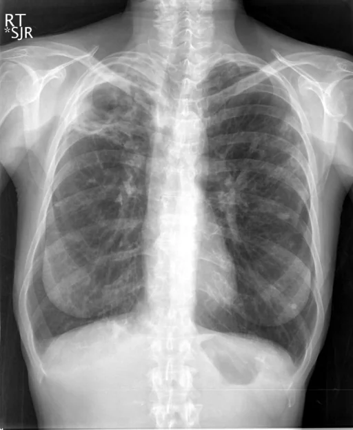
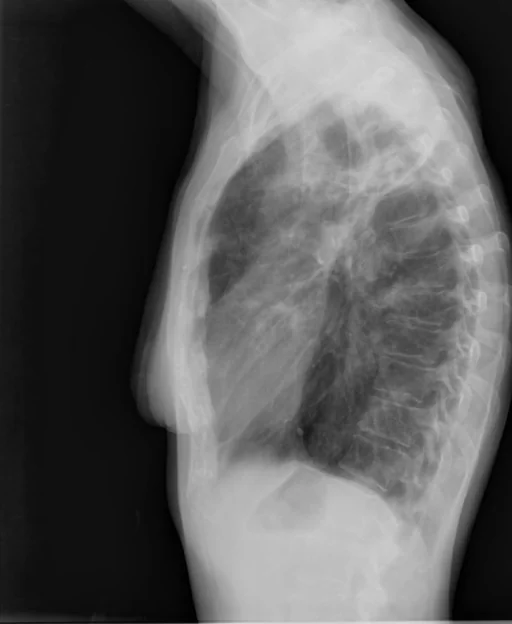
برداشت تشخیصی رادیولوژیست (خلاصهٔ تحلیلی)
یافتههای ثبتشده: کدورت ریه، تغییر حجم ریه، سایر، حفرهسازی (ریه، راس، راست) و ریه (پرهوایی). دستهبندی: «تغییر حجم ریه، کدورت و سایر». وضعیت: «یافتهٔ فعال / نامشخص».
گزارش بالینی اصلی (متن مستند انگلیسی — NoDerivatives)
- اندیکاسیون (Indication)
- Shortness of breath
- یافتهها (Findings)
- There is stable cavity in the right apex, related to prior tuberculosis infection. There is increased opacity in the left upper lobe, peripherally. There is pulmonary hyperexpansion. There is no large effusion or pneumothorax.
- برداشت نهایی (Impression)
- Right upper lobe cavity, consistent with prior tuberculous infection. There is increased right upper lobe opacities compared to the prior study. This could represent reactivation of tuberculosis or other infection.
مشخصات بررسی رادیوگرافی
- شماره پرونده
- CXR ۲۸۱۸
- مودالیتی
- رادیوگرافی قفسهٔ سینه (Chest X-Ray)
- تعداد نماها
- ۲ تصویر
- وضعیت بالینی
- یافتهٔ فعال / نامشخص
کدورتتغییر حجم ریهسایر
این رادیوگرافی قفسهٔ سینه چه یافتهای را نشان میدهد؟
رادیوگرافی قفسهٔ سینه در این پرونده در دستهٔ «تغییر حجم ریه، کدورت و سایر» قرار دارد. یافتهٔ اصلی ثبتشده «کدورت ریه» است و وضعیت مورد «یافتهٔ فعال / نامشخص» است. اطلاعات تکمیلی: سمت: چپ و راست، شدت: پرهوایی.
این متن خلاصهٔ دادههای ساختاریافتهٔ مجموعه است، نه توصیف تصویر.
- محل درگیری: چپ، راست
- شدت و توصیفگران: پرهوایی
- دستهبندی تشخیصی: کدورت، تغییر حجم ریه، سایر
- پوشش تصویربرداری: ۲ نما، رزولوشن اصلی ۵۱۲×۶۲۴ پیکسل
- کلیدواژههای مرتبط: تغییر حجم ریه، Lung Volume (hypoinflation / hyperdistention)، کدورت، Opacity، سایر، Other
کیسهای رادیولوژی مرتبط
منبع، اعتبار علمی و مجوز انتشار
Indiana University Chest X-ray Collection · Open-i / U.S. National Library of Medicine · CC BY-NC-ND 4.0
مجوز انتشار: CC BY-NC-ND 4.0
منبع اصلی داده: https://openi.nlm.nih.gov/