کدورت ریه، بیماری فضای هوایی
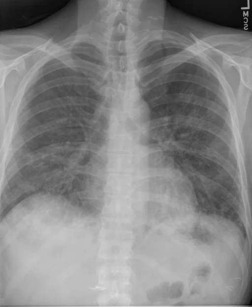
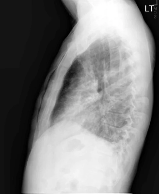
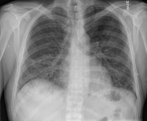
برداشت تشخیصی رادیولوژیست (خلاصهٔ تحلیلی)
یافتههای ثبتشده: کدورت ریه، بیماری فضای هوایی (مزمن)، بیماری فضای هوایی (ریه، دوطرفه) و کدورت (ریه، لوب میانی، دوطرفه، نواری، خفیف). دستهبندی: «بیماری فضای هوایی و کدورت». وضعیت: «ترکیبی (فعال + قدیمی)».
گزارش بالینی اصلی (متن مستند انگلیسی — NoDerivatives)
- اندیکاسیون (Indication)
- Patient for evaluation.
- مقایسه با بررسیهای قبلی (Comparison)
- Single view of the chest.
- یافتهها (Findings)
- Compared to prior examination, there is significant improvement in aeration bilaterally, with improved bilateral airspace opacities. Currently, there are only minimal streaky opacities in the bilateral midlung, which may represent mild residual airspace disease, atelectasis, or underlying changes of chronic lung disease. No large focal consolidations, pneumothorax, or definite pleural effusions identified. The mediastinal silhouette is stable and within normal limits for size and contour. No acute osseous abnormality is identified.
- برداشت نهایی (Impression)
- Significant improvement in bilateral airspace disease and improved aeration bilaterally as described above.
مشخصات بررسی رادیوگرافی
- شماره پرونده
- CXR ۲۳۳۸
- مودالیتی
- رادیوگرافی قفسهٔ سینه (Chest X-Ray)
- تعداد نماها
- ۳ تصویر
- وضعیت بالینی
- ترکیبی (فعال + قدیمی)
کدورتبیماری فضای هوایی
این رادیوگرافی قفسهٔ سینه چه یافتهای را نشان میدهد؟
رادیوگرافی قفسهٔ سینه در این پرونده در دستهٔ «بیماری فضای هوایی و کدورت» قرار دارد. یافتهٔ اصلی ثبتشده «کدورت ریه» است و وضعیت مورد «ترکیبی (فعال + قدیمی)» است. اطلاعات تکمیلی: شدت: دوطرفه و خفیف.
این متن خلاصهٔ دادههای ساختاریافتهٔ مجموعه است، نه توصیف تصویر.
- شدت و توصیفگران: دوطرفه، خفیف، نواری
- دستهبندی تشخیصی: کدورت، بیماری فضای هوایی
- پوشش تصویربرداری: ۳ نما، رزولوشن اصلی ۵۱۲×۶۲۴ پیکسل
- کلیدواژههای مرتبط: بیماری فضای هوایی، Airspace Disease، کدورت، Opacity، ریه، lung
کیسهای رادیولوژی مرتبط
منبع، اعتبار علمی و مجوز انتشار
Indiana University Chest X-ray Collection · Open-i / U.S. National Library of Medicine · CC BY-NC-ND 4.0
مجوز انتشار: CC BY-NC-ND 4.0
منبع اصلی داده: https://openi.nlm.nih.gov/