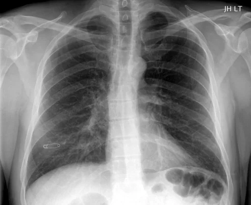
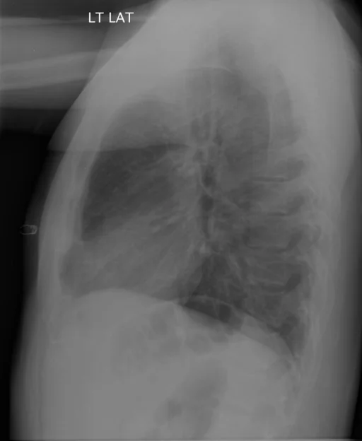
رادیوگرافی قفسهٔ سینه نرمال (طبیعی)
برداشت تشخیصی رادیولوژیست (خلاصهٔ تحلیلی)
مورد بهعنوان «طبیعی» دستهبندی شده است و یافتهٔ بیماری فعالی در فراداده ثبت نشده است.
گزارش بالینی اصلی (متن مستند انگلیسی — NoDerivatives)
- اندیکاسیون (Indication)
- MVA with back pain
- یافتهها (Findings)
- Chest: The cardiomediastinal silhouette is within normal limits for size and contour. The lungs are normally inflated without evidence of focal airspace disease, pleural effusion, or pneumothorax. Thoracic spine: Mild dextro curvature the upper thoracic spine. Evaluation of the upper thoracic bodies is limited secondary to osseous overlap. Vertebral body and disc spaces are maintained. Mild degenerative endplate changes. Lumbar spine: There are 5 nonrib-bearing lumbar type vertebral bodies. Alignment is within normal limits. Vertebral body and disc spaces are maintained. Mild degenerative change without acute displaced fracture or dislocation. Moderate amount of stool.
- برداشت نهایی (Impression)
- Chest. No acute radiographic cardiopulmonary process. Thoracic spine. Mild degenerative change without acute bony abnormality. Lumbar spine. Mild degenerative change without acute bony abnormality.
مشخصات بررسی رادیوگرافی
- شماره پرونده
- CXR ۲۴۸۷
- مودالیتی
- رادیوگرافی قفسهٔ سینه (Chest X-Ray)
- تعداد نماها
- ۲ تصویر
- وضعیت بالینی
- طبیعی
طبیعی
این رادیوگرافی قفسهٔ سینه چه یافتهای را نشان میدهد؟
رادیوگرافی قفسهٔ سینه در این پرونده «طبیعی» دستهبندی شده و یافتهٔ بیماری فعالی در فرادادهٔ آن ثبت نشده است. دستهبندی این پرونده بر پایهٔ برچسبهای ثبتشده در مجموعهٔ داده انجام شده و هیچ برچسب بیماری فعالی به آن اختصاص نیافته است.
این متن خلاصهٔ دادههای ساختاریافتهٔ مجموعه است، نه توصیف تصویر.
- پوشش تصویربرداری: ۲ نما، رزولوشن اصلی ۵۱۲×۴۲۰ پیکسل
- کلیدواژههای مرتبط: طبیعی، Normal، normal، رادیوگرافی طبیعی قفسهٔ سینه، Normal chest radiograph، رادیوگرافی قفسهٔ سینه
کیسهای رادیولوژی مرتبط
منبع، اعتبار علمی و مجوز انتشار
Indiana University Chest X-ray Collection · Open-i / U.S. National Library of Medicine · CC BY-NC-ND 4.0
مجوز انتشار: CC BY-NC-ND 4.0
منبع اصلی داده: https://openi.nlm.nih.gov/