ندول/توده پلور، ضخیمشدگی پلور، اسکار پلور
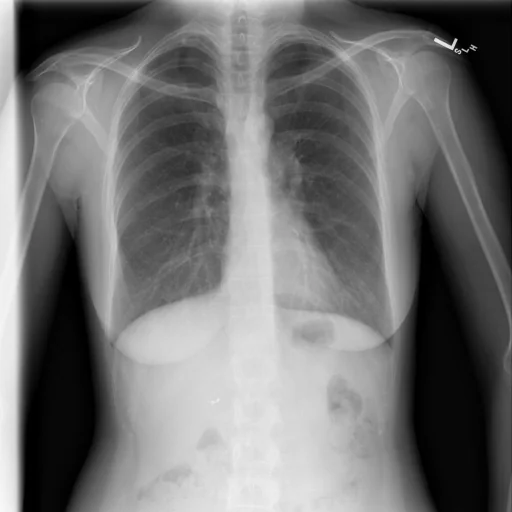
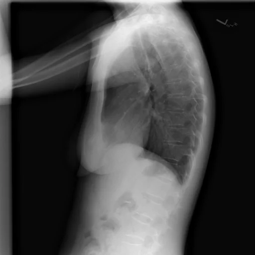
برداشت تشخیصی رادیولوژیست (خلاصهٔ تحلیلی)
یافتههای ثبتشده: ندول/توده پلور، ضخیمشدگی پلور، اسکار پلور، اسکار (پلور، راس، دوطرفه، گِرد) و دفورمیتی (ستون فقرات). دستهبندی: «اسکار (فیبروز موضعی)، دفورمیتی، ندول یا توده و ضخیمشدگی». وضعیت: «یافتهٔ فعال / نامشخص».
گزارش بالینی اصلی (متن مستند انگلیسی — NoDerivatives)
- اندیکاسیون (Indication)
- Adult female. Chest pain after trauma.
- یافتهها (Findings)
- The cardiomediastinal silhouette is normal in size and contour. No focal consolidation, pneumothorax or large pleural effusion. Biapical fibronodular pleural thickening/scarring. There is a step-off deformity of the anterior cortex of the vertebral body (lateral view). Negative for retrosternal density. Prior cholecystectomy. Critical result notification documented through Primordial.
- برداشت نهایی (Impression)
- Buckling deformity of the anterior cortex of the vertebral body. Fracture is possible, if high energy trauma was localized to this region. Correlate with focal tenderness. Chest, if warranted.
مشخصات بررسی رادیوگرافی
- شماره پرونده
- CXR ۳۶۵۸
- مودالیتی
- رادیوگرافی قفسهٔ سینه (Chest X-Ray)
- تعداد نماها
- ۲ تصویر
- وضعیت بالینی
- یافتهٔ فعال / نامشخص
ندول یا تودهضخیمشدگیاسکار (فیبروز موضعی)دفورمیتی
این رادیوگرافی قفسهٔ سینه چه یافتهای را نشان میدهد؟
رادیوگرافی قفسهٔ سینه در این پرونده در دستهٔ «اسکار (فیبروز موضعی)، دفورمیتی، ندول یا توده و ضخیمشدگی» قرار دارد. یافتهٔ اصلی ثبتشده «ندول/توده پلور» است و وضعیت مورد «یافتهٔ فعال / نامشخص» است. اطلاعات تکمیلی: شدت: دوطرفه و گِرد.
این متن خلاصهٔ دادههای ساختاریافتهٔ مجموعه است، نه توصیف تصویر.
- شدت و توصیفگران: دوطرفه، گِرد
- دستهبندی تشخیصی: ندول یا توده، ضخیمشدگی، اسکار (فیبروز موضعی)، دفورمیتی
- پوشش تصویربرداری: ۲ نما، رزولوشن اصلی ۵۱۲×۵۱۲ پیکسل
- کلیدواژههای مرتبط: اسکار (فیبروز موضعی)، Cicatrix، دفورمیتی، Deformity، ندول یا توده، Nodule / Mass
کیسهای رادیولوژی مرتبط
منبع، اعتبار علمی و مجوز انتشار
Indiana University Chest X-ray Collection · Open-i / U.S. National Library of Medicine · CC BY-NC-ND 4.0
مجوز انتشار: CC BY-NC-ND 4.0
منبع اصلی داده: https://openi.nlm.nih.gov/