ندول/توده ریه، آتلکتازی قاعده
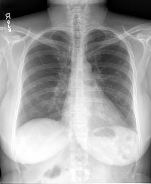
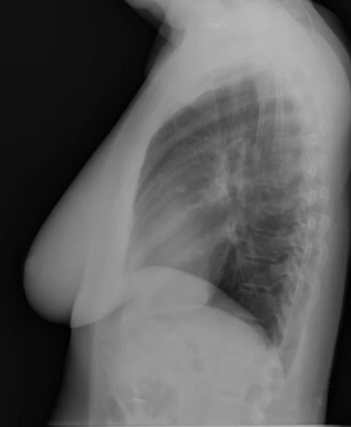
برداشت تشخیصی رادیولوژیست (خلاصهٔ تحلیلی)
یافتههای ثبتشده: ندول/توده ریه، آتلکتازی قاعده، ندول (ریه، لوب میانی، راست، متعدد، کوچک)، ندول (ریه، لوب فوقانی، چپ، متعدد، کوچک) و آتلکتازی ریوی (قاعده، چپ، خفیف). دستهبندی: «آتلکتازی ریه و ندول یا توده». وضعیت: «یافتهٔ فعال / نامشخص».
گزارش بالینی اصلی (متن مستند انگلیسی — NoDerivatives)
- اندیکاسیون (Indication)
- Chest pain
- یافتهها (Findings)
- The cardiac silhouette, upper mediastinum and pulmonary vasculature are within normal limits. There is no acute pulmonary consolidation, large effusion or pneumothorax. There is minimal left basilar atelectasis. There are small bilateral pulmonary nodules measure approximately 5 mm in size in the right midlung and left upper lung fields. These are not well appreciated on the lateral projection.
- برداشت نهایی (Impression)
- 1. No focal pulmonary consolidation or effusion. Minimal left basilar atelectasis. 2. Bilateral pulmonary nodules. These were not present on the prior study and may represent sequela of infection, but could represent neoplastic process. Correlation with history of primary malignancy is recommended. Further evaluation of the thorax could be performed, if clinically indicated.
مشخصات بررسی رادیوگرافی
- شماره پرونده
- CXR ۳۷۵۳
- مودالیتی
- رادیوگرافی قفسهٔ سینه (Chest X-Ray)
- تعداد نماها
- ۲ تصویر
- وضعیت بالینی
- یافتهٔ فعال / نامشخص
ندول یا تودهآتلکتازی ریه
این رادیوگرافی قفسهٔ سینه چه یافتهای را نشان میدهد؟
رادیوگرافی قفسهٔ سینه در این پرونده در دستهٔ «آتلکتازی ریه و ندول یا توده» قرار دارد. یافتهٔ اصلی ثبتشده «ندول/توده ریه» است و وضعیت مورد «یافتهٔ فعال / نامشخص» است. اطلاعات تکمیلی: سمت: چپ و راست، شدت: خفیف و متعدد.
این متن خلاصهٔ دادههای ساختاریافتهٔ مجموعه است، نه توصیف تصویر.
- محل درگیری: چپ، راست
- شدت و توصیفگران: خفیف، متعدد، کوچک
- دستهبندی تشخیصی: ندول یا توده، آتلکتازی ریه
- پوشش تصویربرداری: ۲ نما، رزولوشن اصلی ۵۱۲×۶۲۴ پیکسل
- کلیدواژههای مرتبط: آتلکتازی ریه، Pulmonary Atelectasis، ندول یا توده، Nodule / Mass، ندول، Nodule
کیسهای رادیولوژی مرتبط
منبع، اعتبار علمی و مجوز انتشار
Indiana University Chest X-ray Collection · Open-i / U.S. National Library of Medicine · CC BY-NC-ND 4.0
مجوز انتشار: CC BY-NC-ND 4.0
منبع اصلی داده: https://openi.nlm.nih.gov/