ندول/توده ریه، گرانولوم ریه، کلسیفیکاسیون ریه
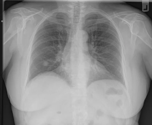
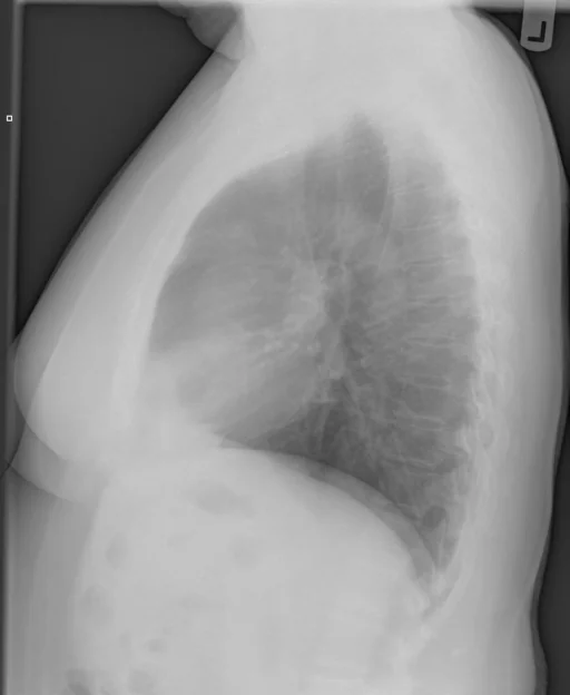
برداشت تشخیصی رادیولوژیست (خلاصهٔ تحلیلی)
یافتههای ثبتشده: ندول/توده ریه، گرانولوم ریه، کلسیفیکاسیون ریه، کلسیفیکاسیون (ریه، ناف ریه، غدد لنفاوی، راست) و گرانولوم (ریه، لوب تحتانی، راست). دستهبندی: «کلسیفیکاسیون، گرانولوم و ندول یا توده». وضعیت: «یافتهٔ فعال / نامشخص».
گزارش بالینی اصلی (متن مستند انگلیسی — NoDerivatives)
- اندیکاسیون (Indication)
- 786.50 pt.states has a soreness lt.lower and lateral rib and upper lt.abd.since having a mammogram one month ago.no or lung complaints./pkd
- مقایسه با بررسیهای قبلی (Comparison)
- INDICATION /
- یافتهها (Findings)
- Chest. Heart size is normal. Pulmonary vasculature is normal. There is a 13 mm nodule in the right lower lobe that is relatively dense, but not obviously calcified on the corresponding rib series. There are probably right hilar calcified lymph nodes. Lungs otherwise are clear. There is no pleural effusion. Left ribs. No fracture or focal bony destruction.
- برداشت نهایی (Impression)
- 1. Chest. Large nodule at the right lung base that probably represents a granuloma although not it is not densely calcified. A low KV P chest radiograph can be obtained for confirmation as a there are no comparison studies available in the. If the patient has an outside chest radiograph, comparison can be and the report addended. 2. Ribs. Normal. Critical result notification documented through Primordial. If there are questions regarding this interpretation, please.
مشخصات بررسی رادیوگرافی
- شماره پرونده
- CXR ۱۸۲۳
- مودالیتی
- رادیوگرافی قفسهٔ سینه (Chest X-Ray)
- تعداد نماها
- ۲ تصویر
- وضعیت بالینی
- یافتهٔ فعال / نامشخص
ندول یا تودهگرانولومکلسیفیکاسیون
این رادیوگرافی قفسهٔ سینه چه یافتهای را نشان میدهد؟
رادیوگرافی قفسهٔ سینه در این پرونده در دستهٔ «کلسیفیکاسیون، گرانولوم و ندول یا توده» قرار دارد. یافتهٔ اصلی ثبتشده «ندول/توده ریه» است و وضعیت مورد «یافتهٔ فعال / نامشخص» است. اطلاعات تکمیلی: سمت: راست.
این متن خلاصهٔ دادههای ساختاریافتهٔ مجموعه است، نه توصیف تصویر.
- محل درگیری: راست
- دستهبندی تشخیصی: ندول یا توده، گرانولوم، کلسیفیکاسیون
- پوشش تصویربرداری: ۲ نما، رزولوشن اصلی ۵۱۲×۴۲۰ پیکسل
- کلیدواژههای مرتبط: کلسیفیکاسیون، Calcinosis، گرانولوم، Granuloma / Granulomatous Disease، ندول یا توده، Nodule / Mass
کیسهای رادیولوژی مرتبط
منبع، اعتبار علمی و مجوز انتشار
Indiana University Chest X-ray Collection · Open-i / U.S. National Library of Medicine · CC BY-NC-ND 4.0
مجوز انتشار: CC BY-NC-ND 4.0
منبع اصلی داده: https://openi.nlm.nih.gov/