ندول/توده ریه، تراکم بافتی (دانسیته)، بزرگی قلب
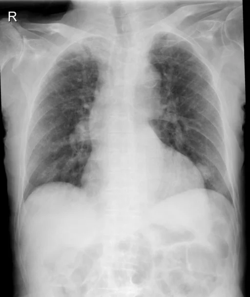
برداشت تشخیصی رادیولوژیست (خلاصهٔ تحلیلی)
یافتههای ثبتشده: ندول/توده ریه، تراکم بافتی (دانسیته)، بزرگی قلب، آئورت (پیچخورده) و آرتریت (ستون فقرات). دستهبندی: «آئورت، آرتریت، بزرگی قلب و تراکم بافتی (دانسیته)». وضعیت: «یافتهٔ فعال / نامشخص».
گزارش بالینی اصلی (متن مستند انگلیسی — NoDerivatives)
- اندیکاسیون (Indication)
- Increasing pain.
- یافتهها (Findings)
- Chest Comparison: There is a 2.6 cm diameter masslike density over the lingula partial obscuration left cardiac device. There may be some ill-defined opacity in the right mid and lower lung zone. No pleural effusion is seen. The heart is borderline enlarged. The aorta is dilated and tortuous. Arthritic changes of the spine are present. Pelvis and left hip There is an impacted and rotated fracture through the neck of the femur on the left. No pelvic fracture is seen. Arthritic changes are present in the lower lumbar spine. Large amount of stool and obscures portions of the pelvis. Femur The femoral images do not the area of the hip fracture. The remaining portions of the femur appear to be intact with no fracture or destructive process. Extensive atherosclerotic vascular disease throughout the superficial femoral artery is present. Left knee There is osteoporosis and mild arthritic changes. No fracture is seen. No dislocation is identified. Severe atherosclerotic changes of the superficial femoral and popliteal artery are seen.
- برداشت نهایی (Impression)
- Chest. 1. Left lower lobe nodule which is worrisome. If there are no prior films available for comparison scan for further evaluation. Pelvis and left hip. Rotated subcapital fracture left hip. Femur. 1. No fracture the remaining portions of the femur. Left knee. 1. Normal for age. Ordering clinician- was called and informed of these critical results.
مشخصات بررسی رادیوگرافی
- شماره پرونده
- CXR ۲۰۶
- مودالیتی
- رادیوگرافی قفسهٔ سینه (Chest X-Ray)
- تعداد نماها
- ۱ تصویر
- وضعیت بالینی
- یافتهٔ فعال / نامشخص
ندول یا تودهتراکم بافتی (دانسیته)بزرگی قلبآئورتآرتریت
این رادیوگرافی قفسهٔ سینه چه یافتهای را نشان میدهد؟
رادیوگرافی قفسهٔ سینه در این پرونده در دستهٔ «آئورت، آرتریت، بزرگی قلب و تراکم بافتی (دانسیته)» قرار دارد. یافتهٔ اصلی ثبتشده «ندول/توده ریه» است و وضعیت مورد «یافتهٔ فعال / نامشخص» است. اطلاعات تکمیلی: سمت: چپ، شدت: مرزی و پیچخورده.
این متن خلاصهٔ دادههای ساختاریافتهٔ مجموعه است، نه توصیف تصویر.
- محل درگیری: چپ
- شدت و توصیفگران: مرزی، پیچخورده
- دستهبندی تشخیصی: ندول یا توده، تراکم بافتی (دانسیته)، بزرگی قلب، آئورت، آرتریت
- پوشش تصویربرداری: ۱ نما، رزولوشن اصلی ۵۱۲×۶۰۸ پیکسل
- کلیدواژههای مرتبط: آئورت، Aorta / Thoracic Aorta، آرتریت، Arthritis، بزرگی قلب، Cardiomegaly
کیسهای رادیولوژی مرتبط
منبع، اعتبار علمی و مجوز انتشار
Indiana University Chest X-ray Collection · Open-i / U.S. National Library of Medicine · CC BY-NC-ND 4.0
مجوز انتشار: CC BY-NC-ND 4.0
منبع اصلی داده: https://openi.nlm.nih.gov/