سایر
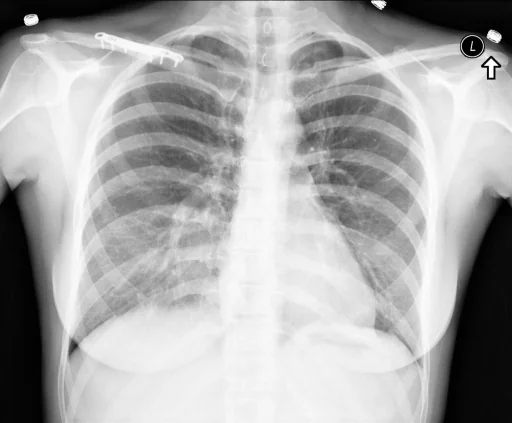
برداشت تشخیصی رادیولوژیست (خلاصهٔ تحلیلی)
یافتههای ثبتشده: سایر و سینهٔ قیفیشکل (خفیف). دستهبندی: «سایر». وضعیت: «یافتهٔ فعال / نامشخص».
گزارش بالینی اصلی (متن مستند انگلیسی — NoDerivatives)
- اندیکاسیون (Indication)
- MVA. Back pain. Right clavicle numbness, with tingling and pain with palpation.
- مقایسه با بررسیهای قبلی (Comparison)
- Chest. Clinical
- یافتهها (Findings)
- Chest: 2 images. Heart size is normal. Mediastinal contours are maintained. There is a mild pectus excavatum deformity. The lungs are clear of focal infiltrate. There is no evidence for pleural effusion or pneumothorax. No convincing acute bony findings. Right shoulder: 3 images. There has been and screw fixation of the midshaft right clavicle. The lateral most screw is fractured. This is age-indeterminate as no prior studies are available for comparison. Otherwise, the surgical appears intact. The humeral head is seen within the glenoid, without evidence for dislocation. No bony fractures are seen. The visualized right ribs appear intact. Right clavicle: 2 images. No clavicle fracture is seen. Once again noted is the surgical fixation, with fracture of the lateral most fixation screw.
- برداشت نهایی (Impression)
- Chest: 1. No acute cardiopulmonary abnormality identified. 2. Pectus excavatum. Right shoulder and right clavicle: 1. No acute bony abnormality identified. 2. The lateral most screw of the clavicular fixation is fractured. This is age-indeterminate as no prior studies are available for comparison. Clinical correlation .
مشخصات بررسی رادیوگرافی
- شماره پرونده
- CXR ۳۲۷۸
- مودالیتی
- رادیوگرافی قفسهٔ سینه (Chest X-Ray)
- تعداد نماها
- ۱ تصویر
- وضعیت بالینی
- یافتهٔ فعال / نامشخص
سایر
این رادیوگرافی قفسهٔ سینه چه یافتهای را نشان میدهد؟
رادیوگرافی قفسهٔ سینه در این پرونده در دستهٔ «سایر» قرار دارد. یافتهٔ اصلی ثبتشده «سینهٔ قیفیشکل (خفیف)» است و وضعیت مورد «یافتهٔ فعال / نامشخص» است. اطلاعات تکمیلی: شدت: خفیف.
این متن خلاصهٔ دادههای ساختاریافتهٔ مجموعه است، نه توصیف تصویر.
- شدت و توصیفگران: خفیف
- دستهبندی تشخیصی: سایر
- پوشش تصویربرداری: ۱ نما، رزولوشن اصلی ۵۱۲×۴۲۳ پیکسل
- کلیدواژههای مرتبط: سایر، Other، سینهٔ قیفیشکل، Funnel Chest، خفیف، mild
کیسهای رادیولوژی مرتبط
منبع، اعتبار علمی و مجوز انتشار
Indiana University Chest X-ray Collection · Open-i / U.S. National Library of Medicine · CC BY-NC-ND 4.0
مجوز انتشار: CC BY-NC-ND 4.0
منبع اصلی داده: https://openi.nlm.nih.gov/