تراکم بافتی (دانسیته)، گرانولوم ریه
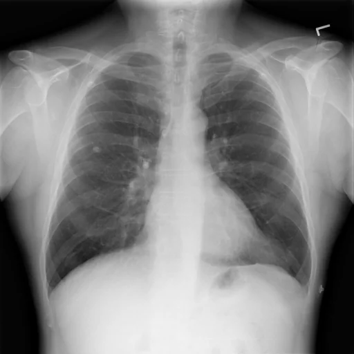
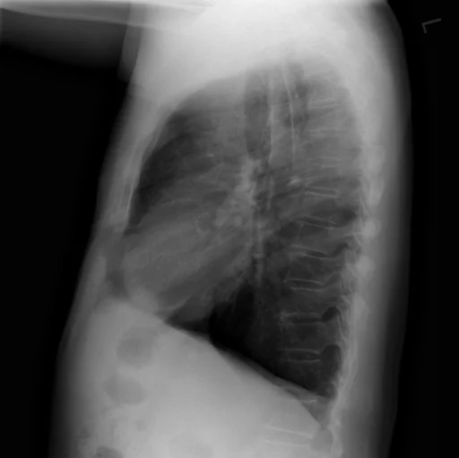
برداشت تشخیصی رادیولوژیست (خلاصهٔ تحلیلی)
یافتههای ثبتشده: تراکم بافتی (دانسیته)، گرانولوم ریه، گرانولوم کلسیفیه (ریه، لوب فوقانی، راست) و تراکم بافتی (دانسیته) (زاویه کاردیوفرنیک، چپ). دستهبندی: «تراکم بافتی (دانسیته) و گرانولوم». وضعیت: «یافتهٔ فعال / نامشخص».
گزارش بالینی اصلی (متن مستند انگلیسی — NoDerivatives)
- اندیکاسیون (Indication)
- Chest pain today. History of stent placement 7+ years ago.
- مقایسه با بررسیهای قبلی (Comparison)
- CT image, CT abdomen which included the lower thorax.
- یافتهها (Findings)
- The radiographic examination consists of frontal and lateral radiographs of the chest. The cardiac silhouette is not enlarged. There has been apparent interval increase in low density convexity at the left cardiophrenic angle. Calcified granuloma is again seen in the right upper lobe. There is no consolidation, pleural effusion or pneumothorax.
- برداشت نهایی (Impression)
- Increased size of density in the left cardiophrenic angle. Primary differential considerations include increased size of prominent epicardial fat, pericardial mass, pleural mass or cardiac aneurysm. CT chest with contrast is recommended. These findings and recommendations were discussed. By ordering clinician telephone. Ordering clinician technologist receipt of the results.
مشخصات بررسی رادیوگرافی
- شماره پرونده
- CXR ۹
- مودالیتی
- رادیوگرافی قفسهٔ سینه (Chest X-Ray)
- تعداد نماها
- ۲ تصویر
- وضعیت بالینی
- یافتهٔ فعال / نامشخص
تراکم بافتی (دانسیته)گرانولوم
این رادیوگرافی قفسهٔ سینه چه یافتهای را نشان میدهد؟
رادیوگرافی قفسهٔ سینه در این پرونده در دستهٔ «تراکم بافتی (دانسیته) و گرانولوم» قرار دارد. یافتهٔ اصلی ثبتشده «تراکم بافتی (دانسیته)» است و وضعیت مورد «یافتهٔ فعال / نامشخص» است. اطلاعات تکمیلی: سمت: چپ و راست.
این متن خلاصهٔ دادههای ساختاریافتهٔ مجموعه است، نه توصیف تصویر.
- محل درگیری: چپ، راست
- دستهبندی تشخیصی: تراکم بافتی (دانسیته)، گرانولوم
- پوشش تصویربرداری: ۲ نما، رزولوشن اصلی ۵۱۲×۵۱۲ پیکسل
- کلیدواژههای مرتبط: تراکم بافتی (دانسیته)، Density، گرانولوم، Granuloma / Granulomatous Disease، گرانولوم کلسیفیه، Calcified Granuloma
کیسهای رادیولوژی مرتبط
منبع، اعتبار علمی و مجوز انتشار
Indiana University Chest X-ray Collection · Open-i / U.S. National Library of Medicine · CC BY-NC-ND 4.0
مجوز انتشار: CC BY-NC-ND 4.0
منبع اصلی داده: https://openi.nlm.nih.gov/