کلسیفیکاسیون مدیاستن
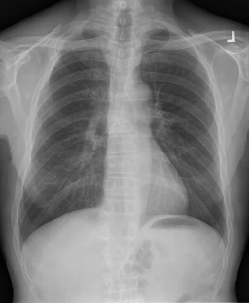
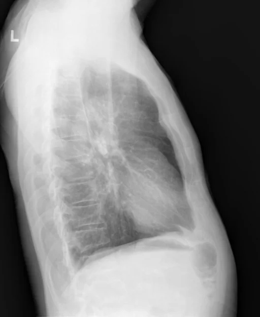
برداشت تشخیصی رادیولوژیست (خلاصهٔ تحلیلی)
یافتههای ثبتشده: کلسیفیکاسیون مدیاستن و کلسیفیکاسیون (مدیاستن، غدد لنفاوی). دستهبندی: «کلسیفیکاسیون». وضعیت: «یافتهٔ فعال / نامشخص».
گزارش بالینی اصلی (متن مستند انگلیسی — NoDerivatives)
- اندیکاسیون (Indication)
- Bilateral hip pain with osteoarthritis. Possible hip replacement.
- یافتهها (Findings)
- On the right there is marked narrowing of the hip joint space uniformly throughout. Osteophyte formation is present with some sclerosis and subchondral cyst formation vertically along the superior acetabulum and femoral head. I do not see evidence for fracture or destructive process. AP view of the femur shows no femoral destructive process or other significant abnormality. For of the Left hip shows near-complete obliteration of the joint space with severe subchondral sclerosis and cystic formation in both the superior acetabulum and superior aspect of the femoral head. No fracture or destructive process is identified. Surgical markers were in the images and left hip for the purpose of surgical planning. PA and lateral chest show the lungs to be clear. There may be some hyperinflation. No pleural effusion is identified. The heart is normal in size. There are calcified mediastinal lymph nodes. The skeletal structures appear normal.
- برداشت نهایی (Impression)
- 1.Severe arthritic changes in both hips left worse than right. 2. Possible COPD but no acute pulmonary disease.
مشخصات بررسی رادیوگرافی
- شماره پرونده
- CXR ۴۵۱
- مودالیتی
- رادیوگرافی قفسهٔ سینه (Chest X-Ray)
- تعداد نماها
- ۲ تصویر
- وضعیت بالینی
- یافتهٔ فعال / نامشخص
کلسیفیکاسیون
این رادیوگرافی قفسهٔ سینه چه یافتهای را نشان میدهد؟
رادیوگرافی قفسهٔ سینه در این پرونده در دستهٔ «کلسیفیکاسیون» قرار دارد. یافتهٔ اصلی ثبتشده «کلسیفیکاسیون مدیاستن» است و وضعیت مورد «یافتهٔ فعال / نامشخص» است.
این متن خلاصهٔ دادههای ساختاریافتهٔ مجموعه است، نه توصیف تصویر.
- دستهبندی تشخیصی: کلسیفیکاسیون
- پوشش تصویربرداری: ۲ نما، رزولوشن اصلی ۵۱۲×۶۲۴ پیکسل
- کلیدواژههای مرتبط: کلسیفیکاسیون، Calcinosis، مدیاستن، mediastinum، غدد لنفاوی، lymph nodes
کیسهای رادیولوژی مرتبط
منبع، اعتبار علمی و مجوز انتشار
Indiana University Chest X-ray Collection · Open-i / U.S. National Library of Medicine · CC BY-NC-ND 4.0
مجوز انتشار: CC BY-NC-ND 4.0
منبع اصلی داده: https://openi.nlm.nih.gov/