کلسیفیکاسیون ریه، دفورمیتی مدیاستن
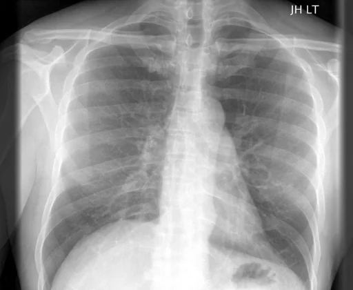
برداشت تشخیصی رادیولوژیست (خلاصهٔ تحلیلی)
یافتههای ثبتشده: کلسیفیکاسیون ریه، دفورمیتی مدیاستن، کلسیفیکاسیون (ریه، ناف ریه، غدد لنفاوی، راست) و دفورمیتی (مدیاستن، خلفی، چپ، کانونی). دستهبندی: «کلسیفیکاسیون و دفورمیتی». وضعیت: «یافتهٔ فعال / نامشخص».
گزارش بالینی اصلی (متن مستند انگلیسی — NoDerivatives)
- یافتهها (Findings)
- PA and lateral views the chest were obtained. The cardiac silhouette is normal in size and configuration. Calcified right hilar lymph nodes. There is focal contour abnormality of the level of the inferior posterior mediastinum, may reflect focal aneurysm of the descending thoracic aorta. The lungs are well aerated. No pneumothorax, pleural effusion, or focal air space consolidation.
- برداشت نهایی (Impression)
- 1. No acute cardiopulmonary disease. 2. Focal convexity of the left inferior posterior mediastinum, may reflect focal aneurysm of the descending thoracic aorta. This has been present and without significant change since. Nonemergent chest CT may be useful.
مشخصات بررسی رادیوگرافی
- شماره پرونده
- CXR ۹۴۹
- مودالیتی
- رادیوگرافی قفسهٔ سینه (Chest X-Ray)
- تعداد نماها
- ۱ تصویر
- وضعیت بالینی
- یافتهٔ فعال / نامشخص
کلسیفیکاسیوندفورمیتی
این رادیوگرافی قفسهٔ سینه چه یافتهای را نشان میدهد؟
رادیوگرافی قفسهٔ سینه در این پرونده در دستهٔ «کلسیفیکاسیون و دفورمیتی» قرار دارد. یافتهٔ اصلی ثبتشده «کلسیفیکاسیون ریه» است و وضعیت مورد «یافتهٔ فعال / نامشخص» است. اطلاعات تکمیلی: سمت: چپ و راست، شدت: کانونی.
این متن خلاصهٔ دادههای ساختاریافتهٔ مجموعه است، نه توصیف تصویر.
- محل درگیری: چپ، راست
- شدت و توصیفگران: کانونی
- دستهبندی تشخیصی: کلسیفیکاسیون، دفورمیتی
- پوشش تصویربرداری: ۱ نما، رزولوشن اصلی ۵۱۲×۴۲۰ پیکسل
- کلیدواژههای مرتبط: کلسیفیکاسیون، Calcinosis، دفورمیتی، Deformity، ریه، lung
کیسهای رادیولوژی مرتبط
منبع، اعتبار علمی و مجوز انتشار
Indiana University Chest X-ray Collection · Open-i / U.S. National Library of Medicine · CC BY-NC-ND 4.0
مجوز انتشار: CC BY-NC-ND 4.0
منبع اصلی داده: https://openi.nlm.nih.gov/